Definition


History
Epidemiology
- The prevalence of glaucoma is for the people who have age 40 to 80 years is 3.54%.
- The prevalence of (Primary Open-Angle Glaucoma) POAG is highest in Africa (4.20%) and the prevalence of Primary Angle-Closure Glaucoma (PACG) is highest in Asia (1.09%)
- In 2013, the number of people (aged 40–80 years) with glaucoma worldwide was estimated to be 64.3 million, increasing to 76.0 million in 2020 and 111.8 million in 2040.
- In the Bayesian meta-regression model, men were more likely to have POAG than women (odds ratio [OR], 1.36; 95% CrI, 1.23–1.52), and after adjusting for age, gender, habitation type, response rate, and year of study, people of African ancestry were more likely to have POAG than people of European ancestry (OR, 2.80; 95% CrI, 1.83–4.06), and people living in urban areas were more likely to have POAG than those in rural areas (OR, 1.58; 95% CrI, 1.19–2.04).
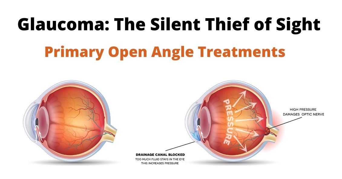
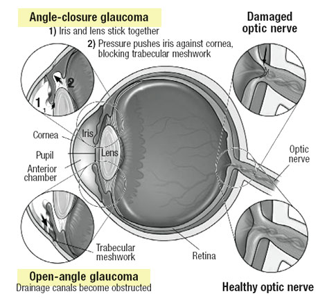
Types

Normal Tension Glaucoma: It is also called as low tension glaucoma. Sometimes with the normal intraocular pressure, optic nerve may get damaged and vision loss may occur. It is due to the poor blood supply to the nerves and thus it causes cell death that carries impulses from the retina to the brain.


CALL OR WHATSAPP : +233271062107
Risk factors
- People above the age of 45
- Family hereditary of glaucoma
- People who with intraocular pressure (IOP)
- Medical conditions such as myopia, diabetes, hyperopia, previous eye injury and long-term use of the corticosteroids
- People of African- American descent have high chances for developing glaucoma than other people
Causes
- The main cause of the glaucoma is a pressure that created in the eye that damages the optic nerve. The normal pressure, which is maintained in our eye ranges between 8 millimeters (mm) and 22 mm of mercury. The eye will be soft when the pressure is low and be harder when the pressure is high.
- The optic nerve is the sensitive part of the eye to high pressures because the nerves easily get damaged by direct pressure or decreased blood flow to the nerve.
- Some gene related to high pressure and optic nerve damage can cause glaucoma
- Some condition that causes glaucoma, such as inflammation of nerves, tumor, diabetes and an advanced cataract
Clinical manifestation of Glaucoma
- Severe eye pain is the first sign of Glaucoma
- Redness in the eye.
- Blurred or cloudy eye, particularly in infants.
- Tunnel vision.
- A headache
- Sick stomach (nausea).
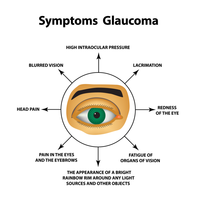

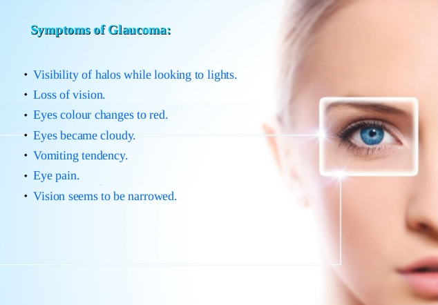
- Rainbow colored halos around any light.
- The blind spot first develops in the side vision (peripheral vision) and outer side of the field of vision.
- In later stages, central vision is affected while seeing is fully lost
- Eye inflammation and infection.
Effects of glaucoma
- One of the major complications of Glaucoma is vision loss.
- Inability to drive because of blurred vision.
- Bleeding and infection of the eye after surgery.
- Most complications are arising after the treatment of Glaucoma.
Diagnosis and Testing of Glaucoma
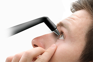
- Intraocular pressure (IOP): IOP is measured using tonometry. During this examination, your eye is numbed using eye drops to rest a lightweight probe gently on the surface of the eyeball. It touches the eyeball and measures the pressure. Sometimes a blast of air is applied to the eye’s surface. The normal IOP should be below 21 mmHg. If your IOP is greater than the 30 mmHg then you are at peak risk of losing vision.
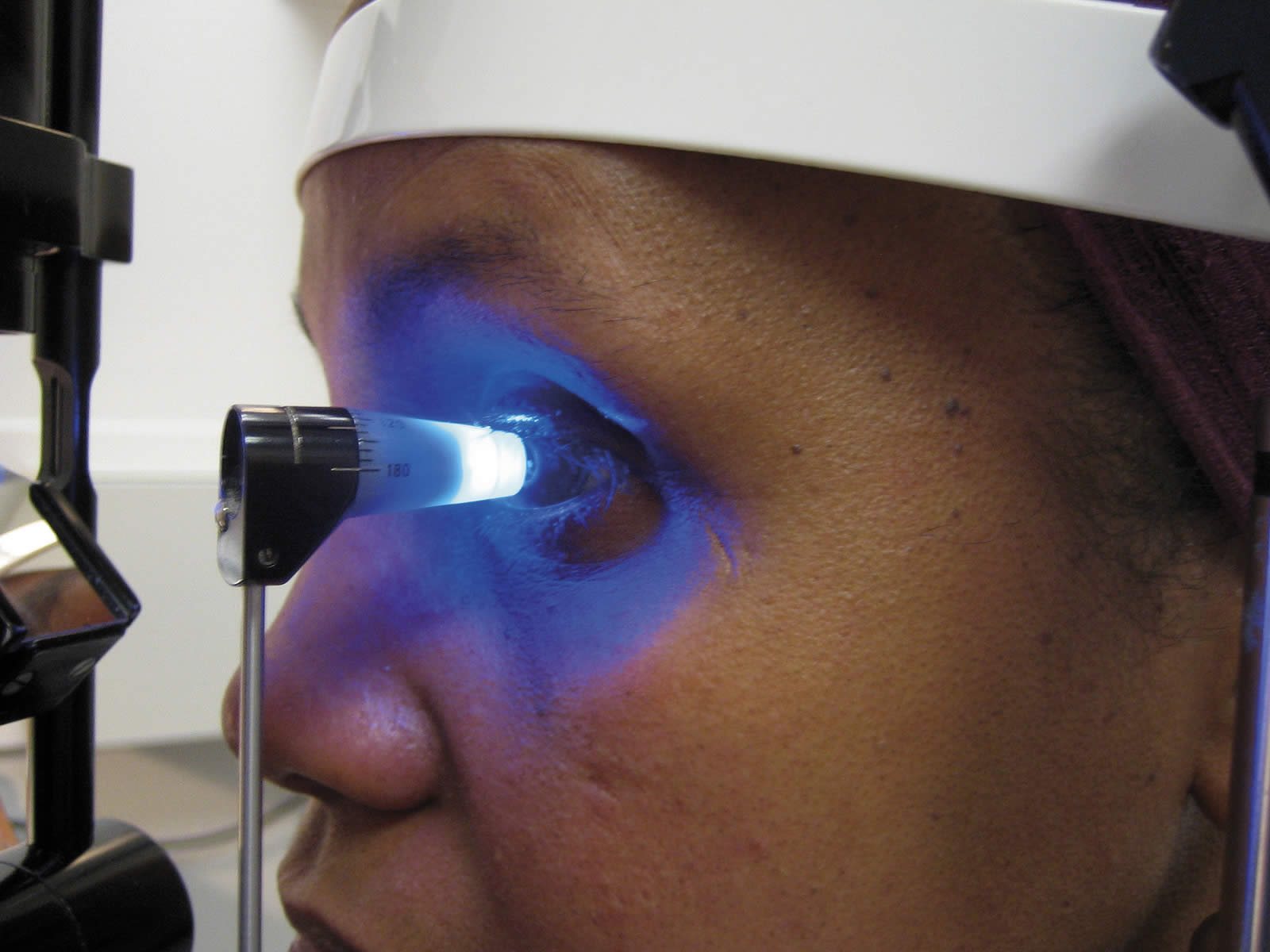

- Assessment of the optic nerve: An optometrist will use eye drops to dilate the pupils and through a slit lamp assess the condition of the optic nerve. A bright light is passed into the eye to visualize the damage in the optic nerve. Eye drops that used to make pupil enlargement may develop difficult to drive and read from a long distance. It is important to have one of your family members along with you to get into the home.
- Visual field test or perimetry: Visual field test finds the missing part of the vision loss. A series of light spots are displayed and asked to look at the spots and will determine the field of vision you have. In which some dots may appear on the sides of eyeballs call peripheral vision.
- Corneal pachymetry: The high corneal thickness may interrupt the values of intraocular pressure and so it is measured using an instrument called pachymetry. It is a device having a pressure sensor probe at its tip is placed on the surface of the eye (Cornea). This procedure takes about 1 to 2 minutes.
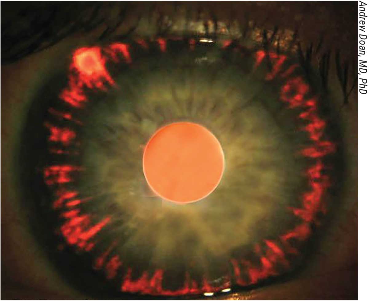
- Gonioscopy: The angle where the cornea and iris meets are measured. During the examination, a hand-held contact lens is placed gently on the eye. This lens has a mirror which will show the angle between the iris and cornea is open or closed.
:max_bytes(150000):strip_icc()/GettyImages-876907828-420c950471f244b2a5868395ebed2b00.jpg)


CALL OR WHATSAPP : +233271062107
Treatment and Medication
Eye drops
Surgery
Prevention of glaucoma
- Regular eye exams should be done
- Daily exercise such as walking, jogging, and running have chances to maintain the normal IOP.
- During yoga practice, you should avoid inverted positions like headstands and shoulder stands.
- Wear protective eyeglasses during your sports activities and home improvement projects.
- If you find any blurred vision immediately go for an eye check-up.
- In general, a check for glaucoma should be done:
- Avoid high amount of salts in your diet if affected with hypersensitive glaucoma.
- If you already have glaucoma, try to avoid coffee and caffeinated beverages to reduce high BP.
- Avoid drinking water at single sip. It is advised to drink a small amount of water during a course of a day.
- Consume very less red wine, green tea, and black chocolate.
- Have foods that are rich in omega 3-fatty acids.
CLICK ON THE WHATSAPP ICON BELOW TO CHAT US DIRECTLY:


We have a Complete Eye Support for the digital age
• Supports healthy vision
• Helps filter blue light from digital devices
• Supports visual processing speed
• Enhances glare recovery time
• Provides all three needed carotenoids specific to inner eye health
•Gluten Free
In today’s digital age, our eyes work harder than ever before. We count on them to filter out UV rays and pollution, but also subject our eyes to hours of artificial blue light coming from mobile devices, computers and televisions every day. Taking care of our eyes is more important than ever to protect from eye strain and long-term damage. And while proper nutrition is the first line of defense, most diets fall far short of the recommended levels of eye-promoting nutrients and antioxidants.
With today’s modern challenges, eye supplements are not only for adults looking to protect their eyes from age-related macular degeneration. Teens and adults of all ages can benefit from our Vision product to support visual processing speed and help replenish macular pigment, enhance glare recovery time and fight exposure to blue light. With the advanced ingredients supplied by our product with Lutemax® 2020, you’ll see clearly why this is the perfect supplement for the digital age.
MORE BENEFITS OF TAKING FOREVER IVISION
1. Improves General Eye Health
2. Improves Clear Vision
3. Improves Conditions In People With Glaucoma and Cataract
4. Contains Vitamin A, Lutemax 2020(containing Lutein and Zeaxanthin) thus nutrients your eyes have been begging for!
5. Stress and Mood Support
6. Cognitive Function
Order Right Here

NATIONWIDE DELIVERY AVAILABLE
CALL OR WHATSAPP: +233271062107
Comments
Post a Comment